Description
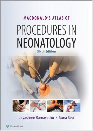
MacDonald’s Atlas of Procedures in Neonatology 6th Edition by Jayashree Ramasethu, ISBN-13: 978-1496394255
[PDF eBook eTextbook] – Available Instantly
- Publisher : LWW
- Publication date : November 29, 2019
- Edition : 6th
- Language : English
- ISBN-10 : 1496394259
- ISBN-13 : 978-1496394255
NOTE: This book is a standalone book and will not include any access codes.
Detailed, step-by-step instructions and abundant full-color illustrations make MacDonald’s Atlas of Procedures in Neonatology, Sixth Edition, an indispensable resource in the neonatal intensive care nursery. This unique reference uses a practical outline format to present clear, easy-to-follow information on indications, preparation, technique, precautions, and how to avoid potential complications. New chapters, new procedural content, and new videos bring you fully up to date with current practice in the NICU.
– Uses a step-by-step format that presents more than 200 photographs and illustrations along with user-friendly procedural descriptions, helping you minimize errors and promote safe practice standards.
– Contains six new chapters: Making Low-Cost Simulation Models for Neonatal Procedures, Delayed Cord Clamping and Cord Milking, Surfactant Administration via Thin Catheter, Amplitude-integrated EEG, VP shunts and EVD Management and Wound Care.
– Includes a new quick-reference appendix with a convenient checklist of procedures.
– Provides access to updated videos that depict both common and emergency neonatal procedures.
Table of Contents:
Cover
Title Page
Copyright
Dedication
Contributors
Video Contributors
Illustration Contributors
Foreword
Preface
Preface to the First Edition
Acknowledgments
Contents
SECTION I Preparation and Support
1 Educational Principles of Simulation-Based Procedural Training
The Need
Definition
The Theory of Simulation-Based Learning
Bloom’s Taxonomy
Kolb’s Experiential Learning Cycle
Procedural Skill Learning
Competency-Based Medical Education and Simulation
A. Identifying and Elucidating the Learning Objectives Specifically Amenable to Simulation
B. Pre-Practice Activities in Preparation for Simulation
C. Choosing the Optimal Simulator (Tables 1.1 to 1.3)
D. A Defined Simulation Environment
E. Pre-Scenario Briefing
F. Running the Appropriately Realistic, Challenging, and Well-Designed Scenario
G. Recording and Identifying the Knowledge and Performance Gaps of the Participants During the Scenario
H. Post-Scenario Debriefing
I. Evaluation of the Simulation Session
Acknowledgements to:
2 Making Low-Cost Simulation Models for Neonatal Procedures
A. Equipment (Additional Model-Specific Equipment Is Listed for Each Model)
B. Chest Tube Model (14)
C. Umbilical Catheter Model
D. Pericardiocentesis Model
E. Suprapubic Bladder Aspiration Model
3 Informed Consent for Procedures
Purpose of Informed Consent
What Are the Requirements for Informed Consent?
Who May Obtain Consent
Types of Informed Consent
What Is Required on a Procedure-Specific Informed Consent Form?
How “Informed” Is Informed Consent?
Special Issues Related to Informed Consent in the Context of Neonates
Consent Refusals
Emergency Procedures
Summary
4 Maintenance of Thermal Homeostasis
A. Definitions
B. Background
C. Indications
D. Equipment, Techniques, and Complications
E. Resource-Limited Settings
F. Special Circumstances/Considerations
5 Methods of Restraint
A. Definitions
B. Indications
C. Contraindications
Restraints Should Not Be Utilized
D. Techniques
Restraints for Procedures/Positioning
Restraints for Vascular Access
E. Precautions
F. Complications
G. Special Considerations
6 Aseptic Preparation
A. Definitions
B. Background
C. Indications
D. Standard Precautions
E. Proper Use of Antiseptics
F. Technique (Video 6.1: Aseptic Preparation)
G. Complications/Precautions
7 Analgesia and Sedation in the Newborn
A. Introduction
B. Definitions
C. General Indications
D. Specific Indications
E. Precautions
F. Advantages and Disadvantages of Commonly Used Agents in the Pediatric Patient
G. Complications
H. Nonpharmacologic Approaches
I. Contraindications
SECTION II Physiologic Monitoring
8 Temperature Monitoring
INTERMITTENT TEMPERATURE MONITORING
A. Equipment
B. Locations
C. Techniques
D. Limitations and Complications
ADDITIONAL INTERMITTENT TEMPERATURE MONITORING
A. Equipment
B. Locations
C. Technique
D. Limitations and Complications
CONTINUOUS TEMPERATURE MONITORING
A. Background
B. Indications
C. Contraindications
D. Equipment Specifications
E. Monitors for Thermistor and Thermocouple Probes
F. Precautions
G. Technique
H. Complications
NEWER ADVANCES IN DEVELOPMENT
A. Thermospot Temperature Indicator
B. Wireless Thermistor Device
C. Wearable Temperature Sensors
References
9 Cardiorespiratory Monitoring
CARDIAC MONITORING
A. Purpose
B. Background
C. Contraindications
D. Limitations
E. Equipment
Hardware—Specifications
Consumables—Specifications
F. Precautions
G. Techniques
H. Complications
RESPIRATORY MONITORING
A. Purpose
B. Background
C. Contraindications
D. Equipment
Hardware—Specifications
Consumables—Specifications
E. Precautions
F. Technique
G. Complications
CARDIORESPIROGRAPH MONITORING
A. Definition
B. Purpose
C. Background
D. Contraindications
E. Equipment
EMERGING TECHNOLOGIES
A. Background
B. Techniques Under Review
C. Implications
10 Blood Pressure Monitoring
NONINVASIVE (INDIRECT) METHODS
AUSCULTATORY MEASUREMENT (MANUAL NONINVASIVE)
A. Background
B. Indications
C. Contraindications
D. Limitations
E. Equipment
F. Precautions (Table 10.1)
G. Technique
H. Complications
OSCILLOMETRIC MEASUREMENT OF ARTERIAL BLOOD PRESSURE (AUTOMATIC NONINVASIVE)
A. Background
B. Indications
C. Contraindications
D. Limitations
E. Equipment
F. Precautions
G. Technique
H. Complications
CONTINUOUS BLOOD PRESSURE MONITORING (INVASIVE)
A. Purpose
B. Background
C. Indications
D. Contraindications
E. Limitations
F. Equipment
G. Technique
H. Complications (Table 10.3)
11 Continuous Blood Gas Monitoring
PULSE OXIMETRY
A. Definitions
B. Background
C. Indications
D. Limitations
E. Equipment
F. Precautions
G. Technique
H. Complications
TRANSCUTANEOUS BLOOD GAS MONITORING
A. Definitions
B. Purpose
C. Background
D. Indications
E. Contraindications
F. Equipment—Specifications
G. Precautions
H. Technique
CONTINUOUS UMBILICAL ARTERY PO2 MONITORING (24,25)
A. Purpose
B. Background
C. Contraindications
D. Equipment
E. Precautions
F. Technique
G. Complications
CONTINUOUS UMBILICAL ARTERY PO2, PCO2, pH, AND TEMPERATURE BLOOD GAS MONITORING (26–32)
A. Purpose
B. Background
C. Contraindications
D. Equipment
E. Precautions
F. Technique
G. Complications
12 End-Tidal Carbon Dioxide Monitoring
CAPNOGRAPHY
A. Definitions
B. Purpose
C. Background
D. Indications
E. Contraindications
F. Limitations (5,18,19)
G. Equipment
H. Precautions
I. Technique
J. Complications
COLORIMETRIC CARBON DIOXIDE MEASUREMENT
A. Indications
B. Procedure
C. Limitations
13 Transcutaneous Bilirubin Monitoring
A. Background
B. Indications
C. Limitations
D. Equipment
E. Special Circumstances/Considerations
F. Techniques
G. Complications
H. Effectiveness
14 Amplitude-Integrated EEG (aEEG)
INDICATIONS FOR aEEG MONITORING
A. Seizures
B. Population At-Risk
C. Equipment
D. Procedure
E. Complications
F. Special Circumstances
INTERPRETATION OF aEEG TRACINGS
A. Background Classification
B. Sleep–Wake Cycling
C. Seizure Recognition
D. Effect of Medications on aEEG
E. Effect of Gestational Age
F. Recognition of Artifacts
G. Limitations
SECTION III Blood Sampling
15 Vessel Localization
TRANSILLUMINATION
A. Indication
B. Contraindications
C. Precautions
D. Equipment
E. Technique
F. Complications
ULTRASONOGRAPHY
A. Background
B. Indication
C. Contraindications
D. Precautions
E. Equipment
F. Technique
G. Complications
NEAR-INFRARED VISUALIZATION
A. Background
B. Indication
C. Contraindications
D. Equipment
E. Technique
16 Venipuncture
A. Indications
B. Contraindications
C. Precautions
D. Special Considerations for Neonates
E. Equipment
F. Technique
General Venipuncture
Drip Technique
Scalp Vein
Proximal Greater Saphenous Vein (7)
External Jugular Vein
G. Complications (8–11)
17 Arterial Puncture
A. Indications (1,2)
B. Contraindications
C. Precautions
D. Selection of Arterial Site
E. Equipment
F. Technique (Video 17.1: Radial Artery Blood Sampling)
General Principles (1,2)
Radial Artery Puncture
Posterior Tibial Puncture
Dorsalis Pedis Puncture
Brachial Artery Puncture
G. Complications (12)
18 Capillary Blood Sampling
A. Purpose
B. Background
C. Indications
D. Contraindications
E. Limitations
F. Equipment
G. Heel-Lancing Devices and Heel Warmers
H. Precautions
I. Technique
J. Specimen Handling
K. Complications
L. Inaccurate Laboratory Results
SECTION IV Miscellaneous Sampling
19 Lumbar Puncture
A. Indications
B. Contraindications
C. Equipment
D. Precautions
E. Technique (Video 19.1: Lumbar Puncture)
F. Complications
20 Subdural Tap
A. Indications (1–7)
B. Contraindications
C. Principles
D. Equipment
E. Technique1
F. Complications
21 Suprapubic Bladder Aspiration
A. Indications (1–8)
B. Contraindications (4,7,8,10)
C. Equipment
D. Precautions
E. Technique
F. Complications
22 Bladder Catheterization
A. Indications (1–4)
B. Contraindications (1,3)
C. Equipment
D. Precautions
E. Technique
Male Infant (1,11,16,17)
Female Infant (1,16–18)
Female Infant in Prone Position (19)
F. Complications
23 Tympanocentesis
A. Indications
B. Contraindications
C. Precautions
D. Technique (7)
E. Complications
24 Bone Marrow Biopsy
A. Definitions
B. Indications
C. Contraindications
D. Precautions
E. Equipment
F. Procedure
G. Special Circumstances
H. Complications1
Acknowledgment
25 Punch Skin Biopsy
A. Definition
B. Indications
C. Types of Skin Biopsy (13)
D. Contraindications
E. Equipment
F. Precautions
G. Technique
H. Complications (13)
26 Ophthalmic Specimen Collection
A. Introduction
B. Indications
C. Relative Contraindications
D. Special Considerations for Ophthalmic Specimen Management
E. Materials
F. Equipment for Identifying Chlamydia and Viral Agents
G. Technique
H. Interpretation of Conjunctival Cytology
I. Complications of Scraping
27 Perimortem Sampling
A. Background
B. Indications (13,14)
C. Discussion With the Family
D. Clinical Information
E. Photographs
F. Examination of the Placenta
G. Perimortem Sampling
H. Imaging: May Be Used Alone or in Conjunction With Autopsy
I. Autopsy
J. Postmortem Family Conference
28 Abdominal Paracentesis
A. Definition
B. Indications
C. Contraindications
D. Equipment
E. Technique (Video 28.1: Abdominal Paracentesis)
F. Complications
SECTION V Vascular Access
29 Peripheral Intravenous Line Placement
A. Indication
B. Equipment
Sterile Equipment (Fig. 29.1)
Nonsterile Clean Equipment
C. Precautions
D. Technique
CONVERSION OF PERIPHERAL IV LINE TO A SALINE LOCK
Technique
Complications
30 Management of Extravasation Injuries
Introduction
A. Assessment
B. Management
1. In All Cases
2. Stage 1 or 2 Extravasation
3. Stage 3 or 4 Extravasation
4. Antidotes and Treatments
5. Wound Management
6. Consultations
31 Umbilical Artery Catheterization
A. Indications
Primary
Secondary
B. Contraindications
C. Equipment
Sterile
Nonsterile
D. Precautions
E. Technique (Video 31.1: Umbilical Vein and Artery Catheterization)
F. Lateral Arteriotomy
G. Umbilical Artery Cutdown
Indications
Contraindications
Equipment
Precautions
Technique (38)
Removal of Catheter
Complications
H. Care of Dwelling Catheter
I. Obtaining Blood Samples From Catheter
Equipment
Technique
J. Removal of UAC
Indications
Technique
K. Complications (48–50)
32 Umbilical Vein Catheterization
A. Indications
B. Contraindications
C. Equipment
D. Precautions
E. Technique (See Video 31.1: Umbilical Vein and Artery Catheterization)
F. Complications
33 Peripheral Arterial Cannulation
A. Indications
B. Contraindications
C. Equipment
Sterile
Non Sterile
Additional Equipment Required for Cut-Down Procedure
Anesthesia/Analgesia
Ultrasound Guided Peripheral Arterial Cannulation
D. Precautions
E. Technique
Standard Technique for Percutaneous Arterial Cannulation
Guidewire-Assisted Radial Artery Cannulation (23)
Radial Artery Cut Down (24)
Posterior Tibial Artery Cannulation by a Cut-Down Procedure
F. Obtaining Arterial Samples
Equipment
Technique I: Three-Drop Method
Technique II: Stopcock Method (a Three-Way Stopcock Needs to Be Interposed Between the Patient and the Transducer)
G. Removal of the Cannula
Indications
Technique
H. Complications of Peripheral Arterial Cannulation
34 Central Venous Catheterization
A. Indications
B. Relative Contraindications
C. General Considerations, Preparation, and Precautions
D. Vessels Amenable to Central Venous Access
E. Position of Catheter Tip (Fig. 34.1)
F. Methods of Vascular Access
G. Types of Central Venous Catheters
Peripherally Inserted Central Venous Catheterization (VIDEO 34.1)
A. Insertion Sites (Fig. 16.1, Table 34.1)
B. Insertion Variations
C. Placement of PICC
D. PICC Dressings (Figs. 34.5G and 34.6)
E. Dressing Changes
F. PICC Care and Maintenance
PLACEMENT OF CENTRAL VENOUS CATHETERS BY SURGICAL CUTDOWN
A. Approach
B. Types of Catheters
C. Contraindications
D. Equipment
Sterile
Nonsterile
E. Techniques
Catheter Placement Via Jugular Veins
Proximal Saphenous Vein Cutdown
F. Sterile Dressing for Surgically Placed Central Venous Lines
Equipment
Precautions
Technique
G. Care of the Catheter When Not in Use for Continuous Infusion
Indications
Equipment
Technique
CATHETER REMOVAL
A. Indications
B. Technique
COMPLICATIONS OF CENTRAL VENOUS CATHETERS (2)
35 Extracorporeal Membrane Oxygenation Cannulation and Decannulation
VENOARTERIAL EXTRACORPOREAL MEMBRANE OXYGENATION—CANNULATION
A. Indications
B. Relative Contraindications for ECMO in the Neonatal Period (5,7)
C. Precautions
D. Personnel, Equipment, and Medications (8)
Personnel
Equipment (Fig. 35.1)
Medications
E. Technique—Preparation for Cannulation
Arterial Cannulation
Venous Cannulation
VENOVENOUS EXTRACORPOREAL MEMBRANE OXYGENATION—CANNULATION
A. Double-Lumen VV Catheters
B. Advantages of VV Bypass
C. Disadvantages of VV Bypass
D. Cannulation Technique
E. Placing Patient on the Extracorporeal Membrane Oxygenation Circuit
F. Closure of the Neck Wound
G. Complications
EXTRACORPOREAL MEMBRANE OXYGENATION—DECANNULATION
A. Indications
B. Contraindications
C. Precautions
D. Personnel, Equipment, and Medications
Personnel
Equipment
Medications
E. Technique
F. Complications
Acknowledgments
36 Management of Vascular Spasm and Thrombosis
A. Definitions
B. Assessment
1. Clinical Diagnosis
2. Diagnostic Imaging
3. Additional Diagnostic Tests
C. Management of Arterial Vascular Spasm/Thromboses
1. Arterial Vascular Spasm
2. Arterial Thromboses (Catheter Related or Idiopathic) (12)
D. Management of Venous Thromboses
1. Catheter-Related Thrombosis
2. Renal Vein Thrombosis (RVT) (4,11,12)
3. Portal Venous Thrombosis (PVT) (13)
E. Anticoagulant/Thrombolytic Therapy
1. General Principles
2. Absolute Contraindications (1,4,14)
3. Relative Contraindications1 (1,4,14)
4. Precautions During Therapy
5. Unfractionated Heparin (UFH)
6. LMWH (16,17)
7. Thrombolytic Agents
8. Complications of Anticoagulation/Fibrinolytic Therapy
F. Surgical Intervention (26)
SECTION VI Respiratory Care
37 Bubble Nasal Continuous Positive Airway Pressure
A. Definition
CPAP Has the Following Physiologic Actions
B. Indications
When to Start b-CPAP?
C. Contraindications
D. Equipment
b-CPAP System Consists of Two Components
E. Technique (See Video 37.1)1
38 Endotracheal Intubation
Introduction
A. Indications
B. Contraindications
C. Considerations
D. Equipment
E. Procedure for Orotracheal Intubation (29)
F. Procedure for Nasotracheal Intubation
G. Procedural Steps for “Push–Pull” Tube Exchange
H. Selective Left Endobronchial Intubation
I. Tracheal Suctioning
J. Intubation Procedural Complications
K. Planned Extubation
L. Placement of Supraglottic/Laryngeal Mask Device
M. Management Considerations for the Patient With an Anticipated Difficult Airway
39 Surfactant Administration via Thin Catheter
A. Definitions
B. Purpose
C. Background
D. Indications
E. Contraindications
F. Precautions
G. Equipment (Fig. 39.1)
H. Technique
Method
I. Special Circumstances
Surfactant Therapy in the Delivery Room
Surfactant Therapy at Gestational Age >32 Weeks
J. Complications
SECTION VII Tube Placement and Care
40 Tracheostomy and Tracheostomy Care
A. Indications
B. Contraindications
C. Precautions
D. Procedure
E. Immediate Postoperative Care (Day 0 Until First Trach Change)
F. Intermediate Postop Care (From First Trach Change Until Transitioned to Home Care)
G. Transitioning to Home Care
H. Complications
41 Thoracostomy
A. Diagnosis of Pneumothorax
A. Equipment (Fig. 41.3)
B. Technique (Video 41.1: Emergency Needle Aspiration)
C. Complications
A. Indications
B. Relative Contraindications
C. Equipment
Sterile
Nonsterile
A. Common Steps
A. Equipment (Fig. 41.6)
B. Procedure
A. Equipment (Fig. 41.9)
B. Procedure
A. Equipment
B. Procedure
A. Equipment
B. Procedure
A. Insertion of Posterior Tube for Fluid Accumulation
B. Post Thoracostomy Tube Placement Steps
A. Preparations
B. Factors Influencing Efficiency of Air Evacuation
C. Removal of Thoracostomy Tube
D. Complications
42 Pericardiocentesis
A. Definitions
B. Background
C. Indications (1,14,20–22)
D. Contraindications
E. Precautions
F. Limitations
G. Equipment
Sterile
Nonsterile (see also H)
H. Procedure (Video 42.1: Pericardiocentesis)
I. Special Circumstances
J. Complications (20–22,26,27)
43 Gastric and Transpyloric Tubes
A. Definitions (1)
ORAL OR NASAL GASTRIC TUBES
A. Indications (2)
B. Types of Tubes
C. Contraindications
D. Precautions
E. Equipment
F. Technique
G. Special Circumstances
H. Complications
TRANSPYLORIC FEEDING TUBE
A. Indications (1,4)
B. Contraindications
C. Precautions (see also Oral or Nasal Gastric Tubes, Part D)
D. Equipment (see also Oral or Nasal Gastric Tubes, Part E)
E. Technique
F. Special Circumstances
G. Complications (see also Oral or Nasal Gastric Tubes, Part H)
44 Gastrostomy
A. Definition
B. Indications
C. Relative Contraindications
D. Preoperative Workup
E. Types of Gastrostomy
F. Postoperative Gastrostomy Care and Maintenance
G. Replacing Gastrostomy Tubes
H. Discontinuation of Gastrostomy (18)
I. Complications (1,18–27)
45 Neonatal Ostomy and Gastrostomy Care
Introduction
A. Definitions
ENTEROSTOMIES AND UROSTOMIES
A. Indications
B. Types of Ostomies
C. Ostomy Assessment
D. Enterostomy Care
E. Equipment
F. Applying the Pouch: Routine/Simple Ostomies (2,6,8)
G. Emptying the Pouch
H. Complicated Stomas and Peristomal Skin Problems (5,9–11)
I. Vesicostomy Care
GASTROSTOMY-JEJUNOSTOMY (G-J) TUBES
A. Indications
B. Types of Tubes
C. Gastrostomy-Jejunostomy Tube Care (8,12)
D. Gastrostomy-Jejunostomy Tube Complications
46 Ventriculoperitoneal Shunt Taps, Percutaneous Ventricular Taps, and External Ventricular Drains
SHUNT TAP
A. Indications
B. Relative Contraindications
C. Equipment (Fig. 46.2)
D. Preprocedure Care
E. Procedure
F. Complications
FONTANELLE TAP/PERCUTANEOUS VENTRICULAR PUNCTURE
A. Indication
B. Preprocedure
C. Procedure
EXTERNAL VENTRICULAR DRAIN
A. Indications
B. Relative Contraindications
C. Equipment
D. Preprocedure Care
E. Procedure
F. Management
G. Monitoring
H. Complications
SECTION VIII Transfusions
47 Delayed Cord Clamping and Cord Milking
A. Definitions
B. Background
C. Factors That Can Either Support or Hinder Placental Transfusion
D. Indications
E. Concerns and Widely Held Beliefs
F. Contraindications
G. Equipment
H. Special Circumstances
I. Technique
J. Complications
48 Transfusion of Blood and Blood Products
OVERVIEW
Blood Products Utilized in Neonates
Sources of Blood Products
A. Precautions (1)
B. Pretransfusion Testing and Processing (1)
C. Equipment
RED BLOOD CELL TRANSFUSIONS
A. Indications
B. Contraindications
C. Technique
FRESH OR RECONSTITUTED WHOLE BLOOD TRANSFUSIONS
A. Indications
B. Precautions (3,4,31)
C. Equipment and Technique
PLATELET TRANSFUSIONS
A. Indications
B. Contraindications (47)
C. Precautions
D. Equipment and Technique
E. Technique for Platelet Administration by Automated Syringe
F. Complications
GRANULOCYTE TRANSFUSIONS
A. Indications
B. Equipment and Technique
C. Precautions
PLASMA PRODUCTS AND CRYOPRECIPITATE
A. Indications (2,54)
B. Contraindications
C. Equipment and Technique
DIRECTED DONOR TRANSFUSIONS
A. Potential Problems
B. Precautions
AUTOLOGOUS FETAL BLOOD TRANSFUSIONS
A. Indications
B. Contraindications
C. Complications
COMPLICATIONS OF BLOOD TRANSFUSIONS
49 Exchange Transfusions
A. Definitions
B. Indications
C. Contraindications
D. Equipment
E. Precautions
F. Preparation for Total or Partial Exchange Transfusion
Blood Product and Volume
Preparation of Infant
Establish Access for ET
Pre-Exchange Laboratory Tests on Infant’s Blood
Prepare Blood
G. Technique (Video 49.1: Exchange Transfusion)
Exchange Transfusion by Push–Pull Technique Through Special Stopcock With Preassembled Tray
Exchange Transfusion Using a Single Umbilical Line and Two Three-Way Stopcocks in Tandem
Exchange Transfusion by Isovolumetric Technique (Central or Peripheral Lines)
H. Postexchange for All Techniques
I. Complications
SECTION IX Miscellaneous Procedures
50 Whole-Body Cooling
A. Indications (2,4) (see Fig. 50.1)
B. Special Circumstances
C. Contraindications
D. Cooling at Birth
E. Securing Rectal or Esophageal Temperature Sensor
Rectal Temperature Sensor
Esophageal Temperature Sensor
F. Supportive Intensive Care With HT
G. Selective Head Cooling
H. Whole-Body Cooling
Passive Cooling
I. Cooling During Transport
Cooling With Adjuncts
Cooling Using a Servo-Controlled Cooling Machine
J. Rewarming
K. Postrewarming Care
L. Complications of Hypothermia
Acknowledgments
51 Removal of Extra Digits and Skin Tags
A. Definitions
B. Indications
C. Precautions
D. Equipment (Fig. 51.2)
E. Procedure or Technique
Polydactyly
Preauricular Tag
F. Complications
52 Neonatal Circumcision
A. Indications
B. Contraindications (4,5)
C. Equipment (7–9)
D. Precautions
E. Techniques
Circumcision With Gomco Circumcision Clamp
Circumcision With Plastibell
Circumcision With Mogen (Crushing) Clamp (Fig. 52.6)
F. Postcircumcision Care
G. Complications
H. Other Complications (Fig. 52.7)
I. Mechanical Device Failures
J. Anesthetic Complications
53 Drainage of Superficial Abscesses
A. Indications
1. Therapeutic
2. Diagnostic
B. Contraindications
C. Principles
D. Equipment
Sterile
Nonsterile
E. Technique
F. Complications
54 Wound Care
A. Definitions
B. Integumentary Assessment Scales
C. Types of Wounds
D. Wound Assessment
E. Wound Cultures
F. Wound Healing
G. Wound Cleansing
H. Wound Dressing Selection
I. Negative Pressure Wound Therapy
J. Diaper Dermatitis
K. Pressure Injuries
L. Debridement
M. Surgical Incisions
N. Congenital Anomalies With Skin Integrity Alterations
O. Ischemic Injuries
55 Phototherapy
A. Indications
B. Contraindications
C. Equipment
Terminology
Devices
D. Technique (Conventional Phototherapy)
Conventional Phototherapy
Fiberoptic Phototherapy
E. Home Phototherapy
F. Efficacy of Phototherapy
G. Discontinuation of Phototherapy and Follow-Up
H. Complications of Phototherapy
56 Intraosseous Infusions
A. Indications
B. Contraindications (6,8,9)
C. Equipment (Fig. 56.1)
Sterile
Optional
Nonsterile
D. Precautions
E. Technique (Video 56.1: Intraosseous Infusion)
Proximal Tibia (Fig. 56.2) (4–6,24)
Distal Tibia (Fig. 56.5) (5,21,24)
Distal Femur (Fig. 56.2) (4)
F. Complications (4,7,30,31)
57 Tapping a Ventricular Reservoir
A. Introduction
B. Indications for Tapping the Reservoir
Based on Ultrasound Measurements
Based on Clinical Symptoms
C. Contraindications
D. Equipment
E. Precautions
F. Technique
G. Successful Tap
H. Follow-Up
I. Complications
58 Treatment of Retinopathy of Prematurity
A. Introduction
B. Screening for ROP
C. Classification of ROP (4)
D. Laser Treatment of ROP (8,9)
E. Intravitreal Injection for ROP
F. Postdischarge Care
G. Outcome
59 Renal Replacement Therapy
Acute Renal Replacement Therapy
Indications
Contraindications
Acute Peritoneal Dialysis
A. Equipment
Sterile
Nonsterile
B. Preprocedure Care
C. Placement of a PD Catheter
D. Management
E. Monitoring
F. Complications
Continuous Renal Replacement Therapy
Prescription
Equipment
Preprocedure Care
Procedure
Management
Monitoring
Complications
60 Neonatal Hearing Screening
A. Purpose
B. Background
C. Indications
D. Types of Hearing Loss
E. Types of Hearing Screen
F. Techniques
G. Specific Protocols
H. Limitations
I. Contraindications
J. Special Circumstances
K. Complications
61 Management of Natal and Neonatal Teeth
Introduction
A. Etiology
B. Clinical Presentation
C. Clinical Assessment
D. Precautions
E. Technique
Nonextraction Case
Extraction Case
F. Complications of Extraction
62 Reducing the Dislocated Newborn Nasal Septum
A. Background
B. Indications
C. Contraindications
D. Equipment
E. Preprocedural Considerations
F. Technique
G. Complications
63 Lingual Frenotomy
A. Definitions
B. Purpose
C. Background
D. Indications
E. Contraindications
F. Limitations
G. Equipment
Sterile
Nonsterile
H. Precautions
I. Technique (1,2,4,10)
J. Complications (1,2,4,11,12)
K. Outcomes (2–4,7–9,13–17)
Appendix A
Appendix B
Appendix C
Appendix D
Appendix E
Index
What makes us different?
• Instant Download
• Always Competitive Pricing
• 100% Privacy
• FREE Sample Available
• 24-7 LIVE Customer Support


