Description
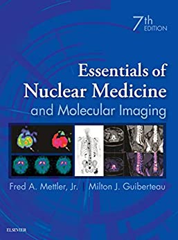
Essentials of Nuclear Medicine and Molecular Imaging 7th Edition by Fred A. Mettler, ISBN-13: 978-0323483193
[PDF eBook eTextbook]
- Publisher: Elsevier; 7th edition (November 21, 2018)
- Language: English
- 560 pages
- ISBN-10: 0323483194
- ISBN-13: 978-0323483193
Covering both the fundamentals and recent developments in this fast-changing field, Essentials of Nuclear Medicine and Molecular Imaging, 7th Edition, is a must-have resource for radiology residents, nuclear medicine residents and fellows, nuclear medicine specialists, and nuclear medicine technicians. Known for its clear and easily understood writing style, superb illustrations, and self-assessment features, this updated classic is an ideal reference for all diagnostic imaging and therapeutic patient care related to nuclear medicine, as well as an excellent review tool for certification or MOC preparation.
- Provides comprehensive, clear explanations of everything from principles of human physiology, pathology, physics, radioactivity, radiopharmaceuticals, radiation safety, and legal requirements to hot topics such as new brain and neuroendocrine tumor agents and hybrid imaging, including PET/MR and PET/CT.
- Covers the imaging of every body system, as well as inflammation, infection and tumor imaging; pearls and pitfalls for every chapter; and pediatric doses and guidelines in compliance with the Image Gently and Image Wisely programs.
- Features a separate self-assessment section on differential diagnoses, imaging procedures and artifacts, and safety issues with unknown cases, questions, answers, and explanations.
- Includes new images and illustrations, for a total of 430 high-quality, multi-modality examples throughout the text.
- Reflects recent advances in the field, including updated nuclear medicine imaging and therapy guidelines • Updated dosimetry values and effective doses for all radiopharmaceuticals with new values from the 2015 International Commission on Radiological Protection • Updated information regarding advances in brain imaging, including amyloid, dopamine transporter and dementia imaging • Inclusion of Ga-68 DOTA PET/CT for neuroendocrine tumors • Expanded information on correlative and hybrid imaging with SPECT/CT • New myocardial agents • and more.
- Contains extensive appendices including updated comprehensive imaging protocols for routine and hybrid imaging, pregnancy and breastfeeding guidelines, pediatric dosages, non-radioactive pharmaceuticals used in interventional and cardiac stress imaging, and radioactivity conversion tables.
Table of Contents:
Cover image
Title Page
Table of Contents
Copyright
Dedication
Preface
Acknowledgments
1 Radioactivity, Radionuclides, and Radiopharmaceuticals
Basic Isotope Notation
Radionuclide Production
Radioactive Decay
Radionuclide Generator Systems
Radionuclides and Radiopharmaceuticals for Imaging
Radiopharmacy Quality Control
Unsealed Radionuclides Used for Therapy
Suggested Readings
2 Instrumentation and Quality Control
Geiger-Mueller Counter
Ionization Chamber
Sodium Iodide Well Counter
Single Probe Counting Systems
Dose Calibrator
Gamma Scintillation Camera
Single-Photon Emission Computed Tomography (SPECT)
SPECT/CT
Positron Emission Tomography (PET)
PET/CT
PET/MRI
Instrumentation Quality Control
Technical Artifacts
Suggested Readings
3 Central Nervous System
Radionuclide Brain Imaging
Cerebrospinal Fluid Imaging
Suggested Readings
4 Thyroid, Parathyroid, and Salivary Glands
Thyroid Radioiodine Uptake and Imaging
Iodine-131 Therapy in Benign Thyroid Disease
Parathyroid Imaging and Localization
Salivary Gland Imaging
Suggested Readings
5 Cardiovascular System
Anatomy and Physiology
SPECT Myocardial Perfusion Imaging
PET Cardiac Imaging
Radionuclide Imaging of Cardiac Function
Suggested Readings
6 Respiratory System
Anatomy and Physiology
Radiopharmaceuticals
Technique
Normal Lung Scan
Clinical Applications
Lung Scanning During Pregnancy
Deep Venous Imaging and Thrombus Detection
Suggested Readings
7 Gastrointestinal Tract
Liver-Spleen Imaging
Hepatic Blood Pool Imaging
Splenic Imaging
Gastrointestinal Bleeding Studies
Meckel Diverticulum Imaging
Hepatobiliary Imaging
Gastroesophageal Function Studies
Abdominal Shunt Evaluation
Suggested Readings
8 Skeletal System
Anatomy and Physiology
Radiopharmaceuticals
Technique
Normal Scan
Clinical Applications
Bone Marrow Imaging
Bone Mineral Measurements
Palliative Therapy of Painful Osseous Metastases
Suggested Readings
9 Genitourinary System and Adrenal Glands
Physiology
Radiopharmaceuticals
Radionuclide Renal Evaluation
Clinical Applications of Renal Imaging
Adrenal Gland Imaging
Suggested Readings
10 Non-PET Neoplasm Imaging and Radionuclide Therapy
Targeted Tumor Imaging
Nonspecific Tumor Imaging
Technetium-99m Sestamibi Tumor Imaging
Lymphoscintigraphy
Radionuclide Tumor Antibody Imaging and Therapy
Suggested Readings
11 Hybrid PET/CT Neoplasm Imaging
18F-FDG PET Imaging
Other PET Radionuclides
Suggested Readings
12 Inflammation and Infection Imaging
Radiolabeled Leukocytes
Gallium Imaging
Fluorine-18 Fluorodeoxyglucose PET/CT Imaging
Future Inflammation Agents
Suggested Readings
13 Authorized User and Radioisotope Safety Issues
Overview
NRC and Other Regulatory Agencies
Types of Licenses
Dose Limits
Radiation Safety Committee
NRC Technical Requirements
Training Required for Use of Unsealed Byproduct Material
Training for Sealed Sources for Diagnosis
Medical Events and Required Reporting
Maintenance of Records
Transportation of Radioactive Materials
Receipt of Radioactive Shipments
Safe Handling and Administration of Radiopharmaceuticals
Generator Breakthrough
PET Radiation Safety
ALARA and Doses to Patients
Release of Individuals After Administration of Radionuclides
Restricted Areas, Radiation Areas, and Signage Posting
Facility Radiation Survey Policies
Waste Disposal
Biological Effects of Ionizing Radiation
Suggested Readings
Self-Evaluation
Unknown Case Sets
Answers to Unknown Case Sets
Answers to Unknown Case Sets
Appendices
Appendix A Characteristics of Radionuclides for Imaging and Therapy
Appendix B.1 Radioactivity Conversion Table for International System (SI) Units (Becquerels to Curies)
Appendix B.2 Radioactivity Conversion Table for International System (SI) Units (Curies to Becquerels)
Appendix C.1 Technetium-99m Decay and Generation Tables
Appendix C.2 Other Radionuclide Decay Tables
Appendix D Injection Techniques and Pediatric Dosages
Appendix E Sample Techniques for Nuclear Imaging
Brain Death or Cerebral Blood Flow Scan
SPECT Brain Perfusion Imaging
Cisternogram
Thyroid Scan (99mTc-Pertechnetate)
Thyroid Scan and Uptake (Iodine-123)
Thyroid Cancer Scan
Parathyroid Scan
Rest Gated Equilibrium Ventriculography
SPECT Myocardial Perfusion Imaging
Exercise Protocol Using a Bruce Multistage or Modified Bruce Treadmill Exercise Protocol
Dipyridamole Pharmacologic Stress Procedure
Adenosine Pharmacologic Stress Procedure
Regadenoson Pharmacologic Stress Procedure
Pulmonary Ventilation Scan (Xenon)
Pulmonary Ventilation Scan (DTPA Aerosol)
Pulmonary Perfusion Scan
Liver and Spleen Scan
SPECT/CT Liver and Spleen Imaging
Hepatobiliary Scan
Meckel Diverticulum Scan
Gastrointestinal Bleeding Scan
Esophageal Transit
Gastroesophageal Reflux/Aspiration
Gastric Emptying
Peritoneovenous (LeVeen) Ascites Shunt Patency
Salivary Gland Imaging
Bone Scan (Technetium-99m)
SPECT Bone Imaging
Bone Marrow Scan
Renal Blood Flow Scan
Renal Scan (A) Cortical Imaging
Renal Scan (B) Glomerular Filtration
Renal Scan (C) Tubular Function
Diuretic (Lasix) Renogram
Captopril Renogram for Diagnosis of Renovascular Hypertension
Radionuclide Cystogram in Children
MIBG Scan for Pheochromocytoma and Neuroblastoma
Gallium Scan for Tumor or Infection
Somatostatin Receptor Scan With Indium-111 Pentetreotide
Lymphoscintigraphy (Sentinel Node Localization)
Molecular Breast Imaging With Breast-Specific Gamma Camera (CZT Detectors)
Leukocyte (White Blood Cell) Scan
PET/CT Tumor Imaging With Fluorine-18 Fluorodeoxyglucose (FDG)
PET Cardiac Imaging With Fluorine-18 Fluorodeoxyglucose (FDG)
PET Brain Imaging With Fluorine-18 Fluorodeoxyglucose (FDG)
PET Amyloid Brain Imaging
PET Cardiac Rest/Stress Imaging With Rubidium-82 or Nitrogen-13 Ammonia
Bone Scan PET/CT (Fluorine-18 Sodium Fluoride)
Red Blood Cell Labeling Techniques
Appendix F Nonradioactive Pharmaceuticals in Nuclear Medicine
Suggested Readings
Appendix G Pregnancy and Breastfeeding
Pregnancy
Breastfeeding
Appendix H.1 General Considerations for Hospitalized Patients Receiving Radionuclide Therapy
Appendix H.2 Special Considerations and Requirements for Iodine-131 Therapy
Nursing Instructions
Release From the Hospital
Appendix I Emergency Procedures for Spills of Radioactive Materials and Special Circumstances
Spills of Radioactive Materials
Minor Spills
Major Spills
Surface Contamination Limits
Emergency Surgery of Patients Who Have Received Therapeutic Amounts of Radionuclides
Emergency Autopsy of Patients Who Have Received Therapeutic Amounts of Radionuclides
Index
Fred A. Mettler, MD, an Elsevier Author, practices diagnostic radiology and nuclear medicine in Albuquerque, where he is Chief of Radiology and Nuclear Medicine at New Mexico Veterans Health Care System. He is currently the U.S. Representative to the United Nations for Radiation Effects.
For more information, visit http://elsevierauthors.com/fredmettlerjr/
What makes us different?
• Instant Download
• Always Competitive Pricing
• 100% Privacy
• FREE Sample Available
• 24-7 LIVE Customer Support

